Skull Anatomy
skull is a strong, bony structure that rests on the neck and encases the brain. It is divided into two main parts: the neurocranium (cranial vault) and the viscerocranium (facial skeleton). The neurocranium surrounds and protects the brain and consists of two components: the skull base, which supports the brain, and the calvaria(skullcap), which covers the brain from above. The viscerocranium primarily supports the facial muscles and various anatomical structures.
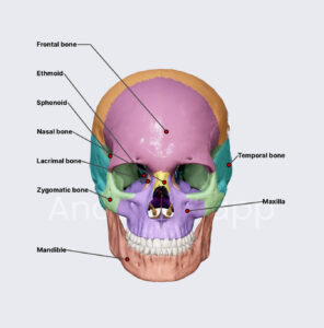
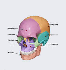
As seen in the skull diagram above, the skull consists of numerous bones—twenty-three in total—some of which are paire
• Ethmoid bone
• Frontal bone
• Inferior nasal concha(e)
• Lacrimal bone(s)
• Mandible
• Maxillary bone(s)
• Nasal bone(s)
• Occipital bone
• Palatine bone(s)
• Parietal bone(s)
• Sphenoid bone
• Temporal bone(s)
• Vomer
• Zygomatic bone(s)
To form a strong and enclosed structure, these bones are joined together by specialized joints called sutures. There are several sutures in the skull, each named after the bones they connect. The most significant ones include the coronal, sagittal, squamous, lambdoid, and palatine sutures, along with key anatomical landmarks such as the lambda, bregma, and pterion.
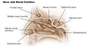
Nose and Nasal Cavity
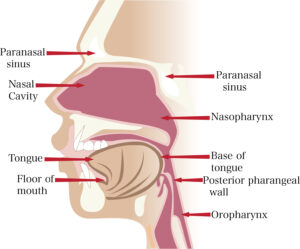
Protruding from the center of your face, the nose is a structure that enables both breathing and the sense of smell. It is composed of nasal bones and cartilage, featuring two openings called nostrils. Located behind the nose is the nasal cavity, which consists of two chambers separated by the nasal septum.
Each cavity contains three curved, shell-like structures known as nasal conchae, beneath which are spaces called nasal meatuses. These meatuses house the nasal openings of various paranasal sinuses, which are situated within the head and skull.
Blood Supply and Innervation
The primary arteries supplying the nose include the:
• Facial artery
• Sphenopalatine artery
• Greater palatine artery
• Ophthalmic artery
The key nerves involved are:
• Olfactory nerve (CN I)– responsible for the sense of smell
• Ophthalmic nerve (CN V1)– a branch of the trigeminal nerve
• Maxillary nerve (CN V2)– another branch of the trigeminal nerve
Sensory innervation of the nose primarily comes from branches of the trigeminal nerve (CN V).
To explore the neurovasculature of the nasal cavity in more detail, check out the study unit below or test your knowledge with a quiz!
The Eye
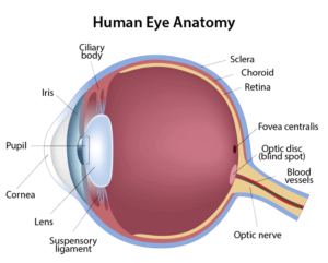
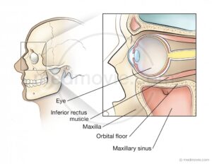
The eyes are two anatomical structures positioned on either side of the nose. Each eye consists of an eyeball suspended within a bony socket in the skull, known as the orbit. The eyeball has a highly intricate and complex anatomy that enables vision. Structurally, it comprises three layers that enclose two jelly-filled compartments, into which the lens is suspended.
Light enters the eye through the pupil, a central black opening controlled by the iris. The pupil regulates the amount of light that reaches the retina, where visual information is processed, ultimately allowing us to see.
Several eye adnexa serve to protect the eyeball and assist in its movement and function. These include:
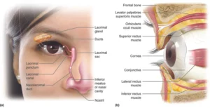
• Eyelids
• Conjunctiva
• Lacrimal apparatus
• Seven extraocular muscles
Blood Supply and Innervation
The ophthalmic artery is the primary blood supply to the eye.
The main nerves involved in eye function include:
• Optic nerve (CN II)– responsible for vision
• Oculomotor nerve (CN III)– controls most eye movements
• Trochlear nerve (CN IV)– controls the superior oblique muscle
• Trigeminal nerve (CN V)– provides sensory innervation
• Abducens nerve (CN VI)– controls the lateral rectus muscle
These nerves reach the eye through three openings located at the back of the orbital cavity.
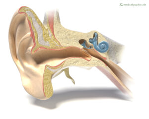
The Ear
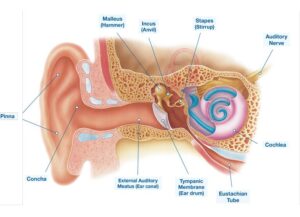
Located on either side of the head, the ears are responsible for hearing and balance. The only visible parts are the auricle and the opening of the ear canal (external auditory canal), while the rest of the ear’s intricate anatomy is hidden within the skull. The ear is made up of three main regions:
• Outer ear– responsible for capturing sound
• Middle ear– transmits sound to the inner ear via the eardrumand three ossicles(malleus, incus, stapes)
• Inner ear– converts sound into nerve impulses through the cochleaand helps maintain balance via the semicircular canals
But the anatomy of the ear extends beyond these regions. Additional structures that surround the ear and assist in its function include the auditory (Eustachian) tube, tegmen tympani, and the labyrinth. The labyrinth also plays a crucial role in maintaining the body’s overall balance.
The primary arteries supplying the ear are the external carotid, maxillary, and basilar arteries, while the main nerves involved are the facial nerve (CN VII) and the vestibulocochlear nerve (CN VIII).
Mouth
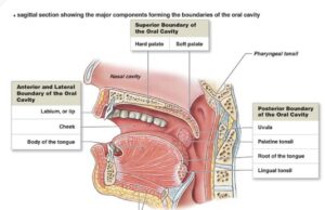
The mouth, anatomically known as the oral cavity, is another prominent facial feature of the head that is not only visible but also involved in speech. It serves as the first part of the digestive system and plays a significant role in the mechanical digestion and mixing of food. The mouth is composed of two main parts:
• Vestibule– the space between the teeth and lips
• Oral cavity proper– the posterior section, which is what most people refer to as the “mouth.”
The oral cavity proper houses several important structures:
• Teeth
• Tongue
• Roof of the mouth(composed of the hard and soft palates)
• Uvula
• Tonsils
• Opening into the oropharynxand its surrounding arches
The most complex and voluminous structure inside the oral cavity is the tongue, which participates in nearly all mouth functions, including chewing, mixing, and swallowing. It consists of two sets of muscles that enable it to move in any direction and take on various shapes within the mouth.
Blood Supply and Innervation
The primary arteries supplying the oral cavity include:
• Descending palatine artery
• Facial artery
• Lingual artery
• Maxillary artery
The main nerves involved in the function of the mouth are:
• Maxillary nerve (CN V2)
• Mandibular nerve (CN V3)
• Vagus nerve (CN X)
• Hypoglossal nerve (CN XII)
• Facial nerve (CN VII)
To learn more about the oral cavity and its structures, check out the following learning materials.
Tooth
Although teeth are part of the oral cavity, their complex anatomy warrants a dedicated section. Throughout a human’s life, two sets of teeth emerge: deciduous teeth, which are shed around six years of age, and permanent teeth, which remain for the rest of the individual’s life. In an adult human, the oral cavity contains thirty-two teeth, arranged into two arches, each consisting of sixteen teeth.
The primary functions of teeth are to bite and mechanically digest food (mastication), but they also play roles in phonation, aesthetics, and nonverbal communication. There are four types of teeth:
• Incisors
• Canines
• Premolars
• Molars
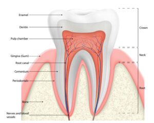
When you open your mouth in front of a mirror, the visible part of each tooth is the crown, which is made of a durable, calcified material for strength. The crown is covered by enameland contains the pulpinside. Each tooth is anchored in the gum (gingiva) by its dental root, the number of which can vary depending on the type of tooth.
Teeth receive their blood supply from the maxillary artery, while their innervation comes from the maxillary nerve (CN V2) and the mandibular nerve (CN V3). To study the complete anatomy of the tooth, check out the following resources.
Neck
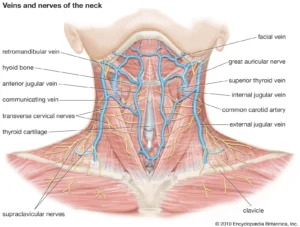
If you thought the previous structures were complex, the neck will definitely impress you. This structure is strong enough to support the head while still being mobile enough to allow it to turn. Externally, the neck is divided into triangles, each of which contains specific muscles, blood vessels, and nerves. Internally, the neck is further divided into compartments, which are separated by various layers of cervical fascia.
To fully understand the triangles and compartments of the neck, explore the following resources and test your knowledge about them.
The anchor point of the neck is the hyoid bone, located at the level of the “Adam’s apple” (laryngeal prominence) in males. Most neck muscles attach to the hyoid, which separates them into two groups: the suprahyoid and infrahyoid muscles. However, other muscles also contribute to the neck’s structure.
Deep within the neck are four major structures: two glands and two passageways:
• Thyroid gland
• Parathyroid glands
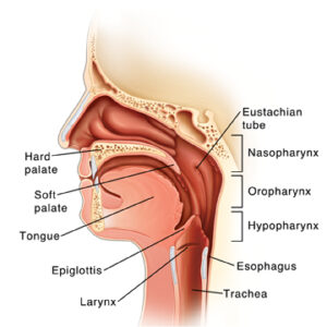
• Pharynx
• Larynx
The thyroid and parathyroid glands are responsible for maintaining the body’s endocrine homeostasis. The pharynxis a muscular passage that connects the nasal and oral cavities to the esophagus and larynx, serving as a conduit for food and air. The larynx, commonly known as the voice box, consists of various cartilages, membranes, ligaments, and muscles. It is essential for producing sound and enabling speech.
The main arteries passing through the neck and supplying it include the common carotid, external carotid, internal carotid, and facial arteries, along with the thyrocervical trunk. The cervical plexus is the primary structure responsible for innervating or passing through the neck.
Since the anatomy of the head and neck is a key topic for anatomy students, we’ve created a custom quiz specifically for this subject. You can tailor the quiz by removing topics or individual structures according to your needs!